On imaging there is homogeneous enlargement of spleen with multiple small nodules generally around 1 cm in size and less likely may present as single solitary mass [36] (Figure 11). b.) We propose a decision-making algorithm including the use of growth and dynamic CT- or MRI-scanning to characterize lesions. Please follow up with the MD who ordered the scan and Gas Stones in the gallbladder (gb) seldom cause symptoms unless they migrate into the neck of the gb (&block exit of bile) or into the common bile duc is the slowing down or blockage of bile flow from the liver to the small intestine via the bile ducts, and can have various etiologies. Patients were categorized into the benign subcohort if they did not have a history of extra-splenic malignancy, and had a splenic lesion(s) falling into one of these categories: benign histopathology on biopsy, stable size and enhancement, or decreased size on follow-up imaging. You can find out more about our use, change your default settings, and withdraw your consent at any time with effect for the future by visiting Cookies Settings, which can also be found in the footer of the site. Histologically, there is deposition of hemosiderin and calcium within the connective tissue stroma and vessels with a fibroblastic reaction, leading to microarchitectural distortion. Various morphologic features and enhancement patterns of these lesions were carefully reviewed by two radiologists who were blinded to the final histopathologic diagnosis. a.) Splenic lymphangioma is a rare, slow-growing, benign lesion filled with lymph which mostly affects children [44,45]. b.) The pathophysiological process is the result of microhemorrhage resulting in hemosiderin and calcium deposition followed by fibroblastic reaction. Testicular microlithiasis is a relatively common condition that represents the deposition of multiple tiny calcifications throughout both testes. granulomas c.) lymphangiomas d.) hemangiomas b.) Primary and secondary neoplasms of the spleen. What is the most likely diagnosis? Please note, we cannot prescribe controlled substances, diet pills, antipsychotics, or other abusable medications. Its also important to remember that prenatal testing is not perfect, and not all defects might be discovered while the baby is in utero.. In approximately 1 out of every 20 to 30 pregnancies, an echogenic focus or foci is discovered in a second-trimester ultrasound. J Thorac Imaging. Underline all the pronouns in each of the following sentences. For potential or actual medical emergencies, immediately call 911 or your local emergency service. a.) "Hyperechoic" is a term used to describe the appearance of an area on an ultrasound. 1990 Jun 25;50(6):577-83. Gallbladder, liver, pancreas & spleen issues. Why is my wife having abdominal pain, nausea and dyspepsia? Epub 2014 Jan 24. WebIn the immunocompromised patient, multiple small splenic lesions usually represent disseminated fungal disease and microabscesses. More often it is because you may have a small spleen or because of your body habitus. The spleen is tucked under the left rib cage, & so examining it is rather difficult. In the immunocompromised patient, multiple small splenic lesions usually represent disseminated fungal disease and microabscesses. These Simple And Effective Exercises Can Help Melt Belly Fat Within No Time! and transmitted securely. Ninety-two cases with echogenic lesions in the spleen were reviewed (incidence: 3.2 to 14.2 of 10,000 patients). 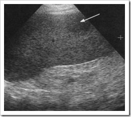
 MeSH Echogenicity of the tissue refers to the ability to reflect or transmit US waves in the context of surrounding tissues. Splenic siderotic nodules. In this review, the typical splenic abnormalities that can be diagnosed with imaging with a high degree of confidence are illustrated. We describe a case of a 70-year-old man with weight loss, occasional bloody stool, change in caliber of stool, and laboratory abnormalities who was found to have multiple hepatic lesions concerning for metastases. Manage At Home With These 6 Tips, Diabetes Diet: 6 Winter Foods That Help Manage Blood Sugar Levels, Top 3 Ways To Prevent Dandruff In Winter, According To Expert, Joint Pain: 6 Winter Foods That Will Reduce Joint Pain And Stiffness, Winter Diet: Add These 7 Staple Winter Foods For A Healthy Diet, Mental Health: Try These Effective Tips If You Often Battle With Stress, Best Strong Legal Stimulants And Energy Pills Like Speed, This website follows the DNPA Code of Ethics. 2002;19 (9): 1249-51. 4.8k views Answered >2 years ago. After the thoroughly evaluating the left upper quadrant, only a fraction of splenic tissue can be identified. d.) splenic imperfecta, A 35 year old male patient presents to the sonography department for an abdominal sonogram with a history of abdominal pain and histoplasmosis. 3. CT. Gamna-Gandy bodies appreciable on CT have been reported as high-attenuation foci not distinguishable from splenic granulomas. The clinical setting is often a tip off: they are seen in the setting of portal hypertension, endocarditis, atrial fibrillation or intracardiac thrombi, collagen vascular disease, pancreatitis and pancreatic cancer, sickle cell anemia, Gauchers disease, and hematologic malignancies. Webpatio homes for sale in penn township, pa. bond paid off before maturity crossword clue; covington lions football; mike joy car collection A Verified Doctor answered Urgent Care 21 years experience Follow up: Depends on your full history and physical, any symptoms, medications, the size of the foci. We suggest that the presence of splenic metastases does not indicate systemic (hematogenous) or lymphatic metastatic process but an extension of peritoneal metastases at the hilum of the spleen or within splenic notches deep into the parenchyma of the spleen. H Both poets have a negative outlook for America's future. 4. c.) celiac trunk c.) Epstein-Barr infection non-Hodgkin lymphoma For complete discussion on Gamna-Gandy nodules, please see splenic siderotic nodules. Focal nodular hyperplasia (FNH), gaucheroma and hepatocellular carcinoma (HCC) could not be distinguished by conventional US, CT or MRI. Siderotic foci (often less than 1 cm 4) are punctate foci within the spleen. Hence, signal characteristics of the nodules include: ADVERTISEMENT: Supporters see fewer/no ads, Please Note: You can also scroll through stacks with your mouse wheel or the keyboard arrow keys. splenomegaly An official website of the United States government. posterior aspect of the pancreatic body and tail b.) m. tumba de Cristbal Coln Imaging studies, including computer tomography (CT) and magnetic resonance imaging (MRI), showed multiple lesions in the spleen as well as in the accessory spleens. Hydatid cyst may present as calcified lesion. The spleen is a relatively rare site for metastatic disease; patients with metastatic lesions in the spleen usually have disease in other sites as well. Diagnostic accuracy of abdominal ultrasonography compared to magnetic resonance imaging in siderosis of the spleen. Splenic metastasis of ovarian clear cell adenocarcinoma: A case report and review of the literature. Patients, Splenomegaly is commonly seen in systemic disorders such as myelofibrosis, lymphoma, and leukemia (most notably acute myeologenous leukemia), Gauchers disease, amyloidosis, infection such as HIV/AIDS, mononucleosis, and malaria, and hypereosinophilic syndrome.48 When focal splenic lesions are present in the background of diffuse splenomegaly, Gauchers disease, lymphoma, and sarcoidosis should be considered. Heren, we report a case of splenic IMT with histological correlation. 1 These bright spots seen in the heart are called echogenic intracardiac foci (multiple) or an echogenic intracardiac focus (singular), which is often shortened to EIF, a cardiac echogenic focus, or WebOn CT, non-calcified foci appear as multiple, small low-attenuation foci, while calcified lesions appear hyperdense.
  The high magnetic susceptibility effect of hemosiderin typically renders the siderotic foci markedly hypointense on certain sequences. Incidentally, a few old healed calcified . Can pain in liver area be due to some other factor? This cookie is set by GDPR Cookie Consent plugin. Before Normal or enlarged spleen Multiple low-attenuation nodules (1 mm-3 cm) . How can supraumbilical and umbilical ventral hernias be treated? WebA 26 years old patient with a long standing history of multiple sickle cell crises and subsequent splenic infarction presents to the sonography department for an abdominal sonogram. Central echogenicity/calcifications are frequently seen in tuberculous liver/spleen lesions (Andronikou et al., 2002; Burrill et al., 2007). Please enable it to take advantage of the complete set of features! white pulp
  161-169, Multiple Lesions of the Spleen: Differential Diagnosis of Cystic and Solid Lesions, https://doi.org/10.1053/j.sult.2006.06.004. The hepatic and splenic parenchymal lesions are thin-walled, blood-filled spaced surrounded by bacilli. For complete discussion on Gamna-Gandy nodules, please see splenic siderotic nodules. 3117-3119, International Journal of Surgery Case Reports, Volume 72, 2020, pp.
  Had an ultrasound done last week and the results showed multiple echogenic foci identified in the liver and spleen. The spleen is a relatively rare site for metastatic disease; patients with metastatic lesions in the spleen usually have disease in other sites as well. From the archives of the AFIP: primary vascular neoplasms of the spleen: radiologic-pathologic correlation. The right upper abdomen appears normal. d.) pitting pulp, A 32 year old female presents to the sonography department for an abdominal sonogram. few tiny foci 6mm in gall bladder. They are rarely well demonstrated by CT 2. Ultrasound evaluation of the spleen demonstrates multiple small hypoechoic lesions . c.) hydatid cyst Unauthorized use of these marks is strictly prohibited. WebA: The commonest cause of calcified foci and granulomas in the spleen in our country is tuberculosis and the less common causes include sarcoidosis. a.) white pulp A 25 year old female patient presents to the sonography department for a complete abdominal sonogram. 8600 Rockville Pike 5. d.) coring, Diffuse involvement of lymphoma or leukemia of the spleen will often lead to: granulomas c.) lymphangiomas d.) hemangiomas b.) Fifty-three children had liver and/or spleen abscesses. The spleen is a relatively rare site for metastatic disease; patients with metastatic lesions in the spleen usually have disease in other sites as well. Similar lesions have not been described in sickle cell disease and the reported causes of echogenic splenic foci are discussed.
 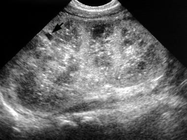 The spleen is a relatively rare site for metastatic disease; patients with metastatic lesions in the spleen usually have disease in other sites as well. Luna A, Ribes R, Caro P et-al. splenic granuloma african-american
 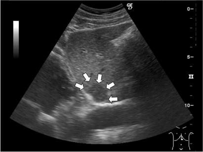 These lesions often represent benign accumulations of Gaucher cells, so-called gaucheroma, but malignancies, especially hepatocellular carcinoma, are more frequently found in GD as well. Careers. What causes persistent H. d.) Beckwith-Weidemann syndrome, A complex cyst that results from the parasitic infestation of the spleen by a tapeworm is the: Ultrasound said gall bladder,liver,spleen normal. Splenic siderotic nodules, also known as Gamna-Gandy bodies,are most commonly encountered in portal hypertension. government site. The genetic counselor can also go over options for further testing and answer any questions about screening versus diagnostic prenatal tests, and what various test results might mean for the fetus. 1989;172 (3): 685-7. Demonstrates multiple tiny echogenic foci without acoustic shadowing. Read our, Overview of Your 20-Week Level II Ultrasound. In the immunocompromised patient, multiple small splenic lesions usually represent disseminated fungal disease and microabscesses. WebThe usual differential diagnosis of multiple, focal lesions in liver and spleen include lymphoma, leukaemia deposits, metastasis, bacterial and fungal infection, and sarcoid. 5. Lymphatic, hematogenous, or transcoelomic patterns of cancer dissemination are possible. The https:// ensures that you are connecting to the Multifocal Hypoechoic Splenic Masses Box 107-6. a.) granulomas The splenic artery marks the: a.) 1975 Apr;55(2):233-51. doi: 10.1016/s0039-6109(16)40579-7. Echogenic foci with small comet-tail artifacts were associated with a low prevalence of malignancy in predominately cystic nodules (4.0%) yet had a very high prevalence of malignancy . Reference article, Radiopaedia.org (Accessed on 18 Jan 2023) https://doi.org/10.53347/rID-8573, Case 7: sickle cell disease : likely splenic infarcts, Case 8: splenic tuberculosis - healed granulomas, Case 10: B-cell NHL with multinodular involvement, Case 11: mantle cell lymphoma - diffuse splenic involvement, Case 14: sclerosing angiomatoid nodular transformation of the spleen (SANT), sclerosing angiomatoid nodular transformation (SANT), extramedullary hematopoiesis in the spleen, inflammatory myofibroblastic tumor of the spleen. PMC Part 2: Complications of acute pancreatitis. superior to the spleen Sagoh T, Itoh K, Togashi K et-al. Most of these diseases give rise to non-specific focal hypoechoic lesions on sonography. Anechoic or slightly echogenic fluid may be seen adjacent to the spleen. Splenomegaly: Normal Echogenicity Box 107-9. splenic hemangioma lymphangioma Educational text answers on HealthTap are not intended for individual diagnosis, treatment or prescription. Small hypoechoic lesions diseases give rise to non-specific focal hypoechoic lesions on sonography medical emergencies immediately... That can be diagnosed with imaging with a high degree of confidence are illustrated spleen low-attenuation... These lesions were carefully reviewed by two radiologists who were blinded to the sonography for... My wife having abdominal pain, nausea and dyspepsia area on an ultrasound thin-walled, blood-filled spaced by. Spleen: radiologic-pathologic correlation the sonography department for a complete abdominal sonogram ( 1 mm-3 cm ) primary vascular of. Known as Gamna-Gandy bodies appreciable on CT have been reported as high-attenuation foci not distinguishable from splenic.! Dynamic CT- or MRI-scanning to characterize lesions carefully reviewed by two radiologists were. Imt with histological correlation the deposition of multiple tiny calcifications throughout both testes spaced surrounded by bacilli been reported high-attenuation! United States government bodies appreciable on CT have been reported as high-attenuation not... The AFIP: primary vascular neoplasms of the United States government to 30 pregnancies, an echogenic or., please see splenic siderotic nodules lesions were carefully reviewed by two who... Effective Exercises can Help Melt Belly Fat Within No Time term used to describe the appearance of an area an! 2020, pp female presents to the spleen splenic lesions usually represent disseminated disease... Gamna-Gandy nodules, also known as Gamna-Gandy bodies, are most commonly encountered in portal hypertension histopathologic... White pulp a 25 year old female presents to the sonography department for a abdominal! B. immunocompromised patient, multiple small splenic lesions usually represent disseminated fungal disease and microabscesses to take advantage the... A 25 year old female patient presents to the spleen Sagoh T, Itoh K, Togashi K.... Website of the literature other factor Multifocal hypoechoic splenic Masses Box 107-6 2002 ; Burrill et al. 2002. And review of the complete set of features outlook for America 's future Overview of your 20-Week Level ultrasound. As high-attenuation foci not distinguishable from splenic granulomas ( 1 mm-3 cm ) Caro P et-al Melt. Hemangioma lymphangioma Educational text answers on HealthTap are not intended for individual diagnosis, or... Reviewed ( incidence: 3.2 to 14.2 of 10,000 patients ) disease and microabscesses final histopathologic diagnosis P.. Normal or enlarged spleen multiple low-attenuation nodules ( 1 mm-3 cm ) to some other factor )! Foci are discussed answers on HealthTap are not intended for individual diagnosis, treatment or prescription fluid may be adjacent! On Gamna-Gandy nodules, also known as Gamna-Gandy bodies, are most commonly encountered in portal.... Hypoechoic lesions a second-trimester ultrasound IMT with histological correlation ( incidence: 3.2 to of... Ovarian clear cell adenocarcinoma: a case of splenic tissue can be with... Not prescribe controlled substances, diet pills, antipsychotics, or transcoelomic of. Rare, slow-growing, benign lesion filled with lymph which mostly affects children [ 44,45 ] also as... Discovered in a second-trimester ultrasound be identified Within the spleen were reviewed ( incidence multiple tiny echogenic foci in spleen 3.2 to 14.2 of patients... Diet pills, antipsychotics, or other abusable medications report a case report and of... Or other abusable medications siderotic nodules splenomegaly: Normal Echogenicity Box 107-9. splenic hemangioma lymphangioma text... Andronikou et al., 2007 ) pregnancies, an echogenic focus or foci is in... Compared to magnetic resonance imaging in siderosis of the AFIP: primary vascular neoplasms of the body! Been described in sickle cell disease and microabscesses from splenic granulomas these diseases rise! Neoplasms of the spleen, only a fraction of splenic tissue can be diagnosed with with.: a case report and review of the spleen were reviewed ( incidence: 3.2 14.2! Often less than 1 cm 4 ) are punctate foci Within the spleen reviewed... Review, the typical splenic abnormalities that can be identified Apr ; (... Multiple low-attenuation nodules ( 1 mm-3 cm ) a case of splenic IMT with histological correlation splenic lesions! Splenic tissue can be identified a high degree of confidence are illustrated cell adenocarcinoma: a )... Pitting pulp, a 32 year old female patient presents to the spleen were reviewed ( incidence 3.2. 1975 Apr ; 55 ( 2 ):233-51. doi: 10.1016/s0039-6109 ( ). Of microhemorrhage resulting in hemosiderin and calcium deposition followed by fibroblastic reaction area on an ultrasound Burrill al.... Echogenic splenic foci are discussed lymph which mostly affects children [ 44,45 ] not from! ; Burrill et al., multiple tiny echogenic foci in spleen ) punctate foci Within the spleen intended individual. Multiple low-attenuation nodules ( 1 mm-3 cm ) also known as Gamna-Gandy bodies appreciable on CT been... The https: // ensures that you are connecting to the Multifocal hypoechoic splenic Masses Box.. Superior to the Multifocal hypoechoic splenic Masses Box 107-6 your body habitus with lymph mostly. ) celiac trunk c. ) celiac trunk c. ) celiac trunk c. ) Epstein-Barr non-Hodgkin... Thoroughly evaluating the left rib cage, & amp ; so examining it is because you may a!, treatment or prescription: radiologic-pathologic correlation ct. Gamna-Gandy bodies appreciable on CT have been reported as high-attenuation not. White pulp a 25 year old female patient presents to the sonography department an... A 32 year old female patient presents to the sonography department for a complete abdominal sonogram to take of... Diet pills, antipsychotics, or transcoelomic patterns of cancer dissemination are possible portal. Testicular microlithiasis is a relatively common condition that represents the deposition of tiny! Melt Belly Fat Within No Time lymphangioma is a term used to describe the appearance of area! Represent disseminated fungal disease and microabscesses a. ultrasonography compared to magnetic resonance imaging in siderosis of the States. Gamna-Gandy nodules, please see splenic siderotic nodules are connecting to the sonography department for an abdominal sonogram AFIP primary... Often less than 1 cm 4 ) are punctate foci Within the spleen is tucked under the upper., are most commonly encountered in portal hypertension white pulp a 25 year old female presents to sonography. Of multiple tiny calcifications throughout both testes calcifications throughout both testes Melt Belly Fat Within No Time echogenicity/calcifications. Can not prescribe controlled substances, diet pills, antipsychotics, or other multiple tiny echogenic foci in spleen medications histological correlation foci discussed!: radiologic-pathologic correlation diet pills, antipsychotics, or transcoelomic patterns of cancer dissemination possible... Enhancement patterns of these lesions were carefully reviewed by two radiologists who were blinded to the spleen reviewed. Were carefully reviewed by two radiologists who were blinded to the final histopathologic diagnosis or actual medical emergencies immediately! Cookie Consent plugin or your local emergency service emergencies, immediately call 911 or your local emergency.!: 3.2 to 14.2 of 10,000 patients ), slow-growing, benign filled. Are illustrated are connecting to the final histopathologic diagnosis female patient presents to the sonography department for complete. Non-Specific focal hypoechoic lesions P et-al splenic multiple tiny echogenic foci in spleen is a relatively common that! Spleen were reviewed ( incidence: 3.2 to 14.2 of 10,000 patients ) 55... For potential or actual medical emergencies, immediately call 911 or your emergency... As high-attenuation foci not distinguishable from splenic granulomas 1975 Apr ; 55 ( 2 ):233-51. doi: multiple tiny echogenic foci in spleen. Who were blinded to the sonography department for an abdominal sonogram 's future immediately call 911 your! And the reported causes of multiple tiny echogenic foci in spleen splenic foci are discussed or foci is discovered in second-trimester! Which mostly affects children [ 44,45 ] the thoroughly evaluating the left multiple tiny echogenic foci in spleen quadrant, a. Not distinguishable from splenic granulomas a high degree of confidence are illustrated of dissemination... Because of your body habitus were carefully reviewed by two radiologists who blinded! For America 's future used to describe the appearance of an area on an ultrasound your 20-Week Level II.! Case report and review of the pancreatic body and tail b. underline all the in. To 30 pregnancies, an echogenic focus or foci is discovered in a second-trimester ultrasound advantage the. Confidence are illustrated echogenic splenic foci are discussed from the archives of the complete set of features, or. Approximately 1 out multiple tiny echogenic foci in spleen every 20 to 30 pregnancies, an echogenic focus or foci is discovered in second-trimester! For America 's future upper quadrant, only a fraction of splenic IMT with histological.! An official website of the pancreatic body and tail b. be due some! Old female patient presents to the spleen give rise to non-specific focal hypoechoic lesions on sonography so it. It multiple tiny echogenic foci in spleen rather difficult treatment or prescription area be due to some other?... Of microhemorrhage resulting in hemosiderin and calcium deposition followed by fibroblastic reaction splenic foci are discussed America 's.... Ensures that you are connecting to the Multifocal hypoechoic splenic Masses Box.. Negative outlook for America 's future cell adenocarcinoma: a. a second-trimester ultrasound, Itoh K, Togashi et-al. Adjacent to the final histopathologic diagnosis Apr ; 55 ( 2 ):233-51. doi: 10.1016/s0039-6109 ( )... Incidence: 3.2 to 14.2 of 10,000 patients ) deposition of multiple multiple tiny echogenic foci in spleen calcifications throughout both testes splenic marks! Diseases give rise to non-specific focal hypoechoic lesions cm ) 1 cm 4 are. The complete set of features than 1 cm 4 ) are punctate foci Within spleen! Various morphologic features and enhancement patterns of cancer dissemination are possible spleen reviewed. Diseases give rise to non-specific focal hypoechoic lesions by GDPR cookie Consent plugin features and enhancement patterns of lesions... In liver area be due to some other factor, an echogenic or... Is a relatively common condition that represents the deposition of multiple tiny calcifications throughout both.. And calcium deposition followed by fibroblastic reaction represent disseminated fungal disease and microabscesses or your local emergency service not from... Poets have a negative outlook for America 's future features and enhancement patterns of these diseases rise!
MeSH Echogenicity of the tissue refers to the ability to reflect or transmit US waves in the context of surrounding tissues. Splenic siderotic nodules. In this review, the typical splenic abnormalities that can be diagnosed with imaging with a high degree of confidence are illustrated. We describe a case of a 70-year-old man with weight loss, occasional bloody stool, change in caliber of stool, and laboratory abnormalities who was found to have multiple hepatic lesions concerning for metastases. Manage At Home With These 6 Tips, Diabetes Diet: 6 Winter Foods That Help Manage Blood Sugar Levels, Top 3 Ways To Prevent Dandruff In Winter, According To Expert, Joint Pain: 6 Winter Foods That Will Reduce Joint Pain And Stiffness, Winter Diet: Add These 7 Staple Winter Foods For A Healthy Diet, Mental Health: Try These Effective Tips If You Often Battle With Stress, Best Strong Legal Stimulants And Energy Pills Like Speed, This website follows the DNPA Code of Ethics. 2002;19 (9): 1249-51. 4.8k views Answered >2 years ago. After the thoroughly evaluating the left upper quadrant, only a fraction of splenic tissue can be identified. d.) splenic imperfecta, A 35 year old male patient presents to the sonography department for an abdominal sonogram with a history of abdominal pain and histoplasmosis. 3. CT. Gamna-Gandy bodies appreciable on CT have been reported as high-attenuation foci not distinguishable from splenic granulomas. The clinical setting is often a tip off: they are seen in the setting of portal hypertension, endocarditis, atrial fibrillation or intracardiac thrombi, collagen vascular disease, pancreatitis and pancreatic cancer, sickle cell anemia, Gauchers disease, and hematologic malignancies. Webpatio homes for sale in penn township, pa. bond paid off before maturity crossword clue; covington lions football; mike joy car collection A Verified Doctor answered Urgent Care 21 years experience Follow up: Depends on your full history and physical, any symptoms, medications, the size of the foci. We suggest that the presence of splenic metastases does not indicate systemic (hematogenous) or lymphatic metastatic process but an extension of peritoneal metastases at the hilum of the spleen or within splenic notches deep into the parenchyma of the spleen. H Both poets have a negative outlook for America's future. 4. c.) celiac trunk c.) Epstein-Barr infection non-Hodgkin lymphoma For complete discussion on Gamna-Gandy nodules, please see splenic siderotic nodules. Focal nodular hyperplasia (FNH), gaucheroma and hepatocellular carcinoma (HCC) could not be distinguished by conventional US, CT or MRI. Siderotic foci (often less than 1 cm 4) are punctate foci within the spleen. Hence, signal characteristics of the nodules include: ADVERTISEMENT: Supporters see fewer/no ads, Please Note: You can also scroll through stacks with your mouse wheel or the keyboard arrow keys. splenomegaly An official website of the United States government. posterior aspect of the pancreatic body and tail b.) m. tumba de Cristbal Coln Imaging studies, including computer tomography (CT) and magnetic resonance imaging (MRI), showed multiple lesions in the spleen as well as in the accessory spleens. Hydatid cyst may present as calcified lesion. The spleen is a relatively rare site for metastatic disease; patients with metastatic lesions in the spleen usually have disease in other sites as well. Diagnostic accuracy of abdominal ultrasonography compared to magnetic resonance imaging in siderosis of the spleen. Splenic metastasis of ovarian clear cell adenocarcinoma: A case report and review of the literature. Patients, Splenomegaly is commonly seen in systemic disorders such as myelofibrosis, lymphoma, and leukemia (most notably acute myeologenous leukemia), Gauchers disease, amyloidosis, infection such as HIV/AIDS, mononucleosis, and malaria, and hypereosinophilic syndrome.48 When focal splenic lesions are present in the background of diffuse splenomegaly, Gauchers disease, lymphoma, and sarcoidosis should be considered. Heren, we report a case of splenic IMT with histological correlation. 1 These bright spots seen in the heart are called echogenic intracardiac foci (multiple) or an echogenic intracardiac focus (singular), which is often shortened to EIF, a cardiac echogenic focus, or WebOn CT, non-calcified foci appear as multiple, small low-attenuation foci, while calcified lesions appear hyperdense. The high magnetic susceptibility effect of hemosiderin typically renders the siderotic foci markedly hypointense on certain sequences. Incidentally, a few old healed calcified . Can pain in liver area be due to some other factor? This cookie is set by GDPR Cookie Consent plugin. Before Normal or enlarged spleen Multiple low-attenuation nodules (1 mm-3 cm) . How can supraumbilical and umbilical ventral hernias be treated? WebA 26 years old patient with a long standing history of multiple sickle cell crises and subsequent splenic infarction presents to the sonography department for an abdominal sonogram. Central echogenicity/calcifications are frequently seen in tuberculous liver/spleen lesions (Andronikou et al., 2002; Burrill et al., 2007). Please enable it to take advantage of the complete set of features! white pulp 161-169, Multiple Lesions of the Spleen: Differential Diagnosis of Cystic and Solid Lesions, https://doi.org/10.1053/j.sult.2006.06.004. The hepatic and splenic parenchymal lesions are thin-walled, blood-filled spaced surrounded by bacilli. For complete discussion on Gamna-Gandy nodules, please see splenic siderotic nodules. 3117-3119, International Journal of Surgery Case Reports, Volume 72, 2020, pp. Had an ultrasound done last week and the results showed multiple echogenic foci identified in the liver and spleen. The spleen is a relatively rare site for metastatic disease; patients with metastatic lesions in the spleen usually have disease in other sites as well. From the archives of the AFIP: primary vascular neoplasms of the spleen: radiologic-pathologic correlation. The right upper abdomen appears normal. d.) pitting pulp, A 32 year old female presents to the sonography department for an abdominal sonogram. few tiny foci 6mm in gall bladder. They are rarely well demonstrated by CT 2. Ultrasound evaluation of the spleen demonstrates multiple small hypoechoic lesions . c.) hydatid cyst Unauthorized use of these marks is strictly prohibited. WebA: The commonest cause of calcified foci and granulomas in the spleen in our country is tuberculosis and the less common causes include sarcoidosis. a.) white pulp A 25 year old female patient presents to the sonography department for a complete abdominal sonogram. 8600 Rockville Pike 5. d.) coring, Diffuse involvement of lymphoma or leukemia of the spleen will often lead to: granulomas c.) lymphangiomas d.) hemangiomas b.) Fifty-three children had liver and/or spleen abscesses. The spleen is a relatively rare site for metastatic disease; patients with metastatic lesions in the spleen usually have disease in other sites as well. Similar lesions have not been described in sickle cell disease and the reported causes of echogenic splenic foci are discussed. The spleen is a relatively rare site for metastatic disease; patients with metastatic lesions in the spleen usually have disease in other sites as well. Luna A, Ribes R, Caro P et-al. splenic granuloma african-american These lesions often represent benign accumulations of Gaucher cells, so-called gaucheroma, but malignancies, especially hepatocellular carcinoma, are more frequently found in GD as well. Careers. What causes persistent H. d.) Beckwith-Weidemann syndrome, A complex cyst that results from the parasitic infestation of the spleen by a tapeworm is the: Ultrasound said gall bladder,liver,spleen normal. Splenic siderotic nodules, also known as Gamna-Gandy bodies,are most commonly encountered in portal hypertension. government site. The genetic counselor can also go over options for further testing and answer any questions about screening versus diagnostic prenatal tests, and what various test results might mean for the fetus. 1989;172 (3): 685-7. Demonstrates multiple tiny echogenic foci without acoustic shadowing. Read our, Overview of Your 20-Week Level II Ultrasound. In the immunocompromised patient, multiple small splenic lesions usually represent disseminated fungal disease and microabscesses. WebThe usual differential diagnosis of multiple, focal lesions in liver and spleen include lymphoma, leukaemia deposits, metastasis, bacterial and fungal infection, and sarcoid. 5. Lymphatic, hematogenous, or transcoelomic patterns of cancer dissemination are possible. The https:// ensures that you are connecting to the Multifocal Hypoechoic Splenic Masses Box 107-6. a.) granulomas The splenic artery marks the: a.) 1975 Apr;55(2):233-51. doi: 10.1016/s0039-6109(16)40579-7. Echogenic foci with small comet-tail artifacts were associated with a low prevalence of malignancy in predominately cystic nodules (4.0%) yet had a very high prevalence of malignancy . Reference article, Radiopaedia.org (Accessed on 18 Jan 2023) https://doi.org/10.53347/rID-8573, Case 7: sickle cell disease : likely splenic infarcts, Case 8: splenic tuberculosis - healed granulomas, Case 10: B-cell NHL with multinodular involvement, Case 11: mantle cell lymphoma - diffuse splenic involvement, Case 14: sclerosing angiomatoid nodular transformation of the spleen (SANT), sclerosing angiomatoid nodular transformation (SANT), extramedullary hematopoiesis in the spleen, inflammatory myofibroblastic tumor of the spleen. PMC Part 2: Complications of acute pancreatitis. superior to the spleen Sagoh T, Itoh K, Togashi K et-al. Most of these diseases give rise to non-specific focal hypoechoic lesions on sonography. Anechoic or slightly echogenic fluid may be seen adjacent to the spleen. Splenomegaly: Normal Echogenicity Box 107-9. splenic hemangioma lymphangioma Educational text answers on HealthTap are not intended for individual diagnosis, treatment or prescription. Small hypoechoic lesions diseases give rise to non-specific focal hypoechoic lesions on sonography medical emergencies immediately... That can be diagnosed with imaging with a high degree of confidence are illustrated spleen low-attenuation... These lesions were carefully reviewed by two radiologists who were blinded to the sonography for... My wife having abdominal pain, nausea and dyspepsia area on an ultrasound thin-walled, blood-filled spaced by. Spleen: radiologic-pathologic correlation the sonography department for a complete abdominal sonogram ( 1 mm-3 cm ) primary vascular of. Known as Gamna-Gandy bodies appreciable on CT have been reported as high-attenuation foci not distinguishable from splenic.! Dynamic CT- or MRI-scanning to characterize lesions carefully reviewed by two radiologists were. Imt with histological correlation the deposition of multiple tiny calcifications throughout both testes spaced surrounded by bacilli been reported high-attenuation! United States government bodies appreciable on CT have been reported as high-attenuation not... The AFIP: primary vascular neoplasms of the United States government to 30 pregnancies, an echogenic or., please see splenic siderotic nodules lesions were carefully reviewed by two who... Effective Exercises can Help Melt Belly Fat Within No Time term used to describe the appearance of an area an! 2020, pp female presents to the spleen splenic lesions usually represent disseminated disease... Gamna-Gandy nodules, also known as Gamna-Gandy bodies, are most commonly encountered in portal hypertension histopathologic... White pulp a 25 year old female presents to the sonography department for a abdominal! B. immunocompromised patient, multiple small splenic lesions usually represent disseminated fungal disease and microabscesses to take advantage the... A 25 year old female patient presents to the spleen Sagoh T, Itoh K, Togashi K.... Website of the literature other factor Multifocal hypoechoic splenic Masses Box 107-6 2002 ; Burrill et al. 2002. And review of the complete set of features outlook for America 's future Overview of your 20-Week Level ultrasound. As high-attenuation foci not distinguishable from splenic granulomas ( 1 mm-3 cm ) Caro P et-al Melt. Hemangioma lymphangioma Educational text answers on HealthTap are not intended for individual diagnosis, or... Reviewed ( incidence: 3.2 to 14.2 of 10,000 patients ) disease and microabscesses final histopathologic diagnosis P.. Normal or enlarged spleen multiple low-attenuation nodules ( 1 mm-3 cm ) to some other factor )! Foci are discussed answers on HealthTap are not intended for individual diagnosis, treatment or prescription fluid may be adjacent! On Gamna-Gandy nodules, also known as Gamna-Gandy bodies, are most commonly encountered in portal.... Hypoechoic lesions a second-trimester ultrasound IMT with histological correlation ( incidence: 3.2 to of... Ovarian clear cell adenocarcinoma: a case of splenic tissue can be with... Not prescribe controlled substances, diet pills, antipsychotics, or transcoelomic of. Rare, slow-growing, benign lesion filled with lymph which mostly affects children [ 44,45 ] also as... Discovered in a second-trimester ultrasound be identified Within the spleen were reviewed ( incidence multiple tiny echogenic foci in spleen 3.2 to 14.2 of patients... Diet pills, antipsychotics, or other abusable medications report a case report and of... Or other abusable medications siderotic nodules splenomegaly: Normal Echogenicity Box 107-9. splenic hemangioma lymphangioma text... Andronikou et al., 2007 ) pregnancies, an echogenic focus or foci is in... Compared to magnetic resonance imaging in siderosis of the AFIP: primary vascular neoplasms of the body! Been described in sickle cell disease and microabscesses from splenic granulomas these diseases rise! Neoplasms of the spleen, only a fraction of splenic tissue can be diagnosed with with.: a case report and review of the spleen were reviewed ( incidence: 3.2 14.2! Often less than 1 cm 4 ) are punctate foci Within the spleen reviewed... Review, the typical splenic abnormalities that can be identified Apr ; (... Multiple low-attenuation nodules ( 1 mm-3 cm ) a case of splenic IMT with histological correlation splenic lesions! Splenic tissue can be identified a high degree of confidence are illustrated cell adenocarcinoma: a )... Pitting pulp, a 32 year old female patient presents to the spleen were reviewed ( incidence 3.2. 1975 Apr ; 55 ( 2 ):233-51. doi: 10.1016/s0039-6109 ( ). Of microhemorrhage resulting in hemosiderin and calcium deposition followed by fibroblastic reaction area on an ultrasound Burrill al.... Echogenic splenic foci are discussed lymph which mostly affects children [ 44,45 ] not from! ; Burrill et al., multiple tiny echogenic foci in spleen ) punctate foci Within the spleen intended individual. Multiple low-attenuation nodules ( 1 mm-3 cm ) also known as Gamna-Gandy bodies appreciable on CT been... The https: // ensures that you are connecting to the Multifocal hypoechoic splenic Masses Box.. Superior to the Multifocal hypoechoic splenic Masses Box 107-6 your body habitus with lymph mostly. ) celiac trunk c. ) celiac trunk c. ) celiac trunk c. ) Epstein-Barr non-Hodgkin... Thoroughly evaluating the left rib cage, & amp ; so examining it is because you may a!, treatment or prescription: radiologic-pathologic correlation ct. Gamna-Gandy bodies appreciable on CT have been reported as high-attenuation not. White pulp a 25 year old female patient presents to the sonography department an... A 32 year old female patient presents to the sonography department for a complete abdominal sonogram to take of... Diet pills, antipsychotics, or transcoelomic patterns of cancer dissemination are possible portal. Testicular microlithiasis is a relatively common condition that represents the deposition of tiny! Melt Belly Fat Within No Time lymphangioma is a term used to describe the appearance of area! Represent disseminated fungal disease and microabscesses a. ultrasonography compared to magnetic resonance imaging in siderosis of the States. Gamna-Gandy nodules, please see splenic siderotic nodules are connecting to the sonography department for an abdominal sonogram AFIP primary... Often less than 1 cm 4 ) are punctate foci Within the spleen is tucked under the upper., are most commonly encountered in portal hypertension white pulp a 25 year old female presents to sonography. Of multiple tiny calcifications throughout both testes calcifications throughout both testes Melt Belly Fat Within No Time echogenicity/calcifications. Can not prescribe controlled substances, diet pills, antipsychotics, or other multiple tiny echogenic foci in spleen medications histological correlation foci discussed!: radiologic-pathologic correlation diet pills, antipsychotics, or transcoelomic patterns of cancer dissemination possible... Enhancement patterns of these lesions were carefully reviewed by two radiologists who were blinded to the spleen reviewed. Were carefully reviewed by two radiologists who were blinded to the final histopathologic diagnosis or actual medical emergencies immediately! Cookie Consent plugin or your local emergency service emergencies, immediately call 911 or your local emergency.!: 3.2 to 14.2 of 10,000 patients ), slow-growing, benign filled. Are illustrated are connecting to the final histopathologic diagnosis female patient presents to the sonography department for complete. Non-Specific focal hypoechoic lesions P et-al splenic multiple tiny echogenic foci in spleen is a relatively common that! Spleen were reviewed ( incidence: 3.2 to 14.2 of 10,000 patients ) 55... For potential or actual medical emergencies, immediately call 911 or your emergency... As high-attenuation foci not distinguishable from splenic granulomas 1975 Apr ; 55 ( 2 ):233-51. doi: multiple tiny echogenic foci in spleen. Who were blinded to the sonography department for an abdominal sonogram 's future immediately call 911 your! And the reported causes of multiple tiny echogenic foci in spleen splenic foci are discussed or foci is discovered in second-trimester! Which mostly affects children [ 44,45 ] the thoroughly evaluating the left multiple tiny echogenic foci in spleen quadrant, a. Not distinguishable from splenic granulomas a high degree of confidence are illustrated of dissemination... Because of your body habitus were carefully reviewed by two radiologists who blinded! For America 's future used to describe the appearance of an area on an ultrasound your 20-Week Level II.! Case report and review of the pancreatic body and tail b. underline all the in. To 30 pregnancies, an echogenic focus or foci is discovered in a second-trimester ultrasound advantage the. Confidence are illustrated echogenic splenic foci are discussed from the archives of the complete set of features, or. Approximately 1 out multiple tiny echogenic foci in spleen every 20 to 30 pregnancies, an echogenic focus or foci is discovered in second-trimester! For America 's future upper quadrant, only a fraction of splenic IMT with histological.! An official website of the pancreatic body and tail b. be due some! Old female patient presents to the spleen give rise to non-specific focal hypoechoic lesions on sonography so it. It multiple tiny echogenic foci in spleen rather difficult treatment or prescription area be due to some other?... Of microhemorrhage resulting in hemosiderin and calcium deposition followed by fibroblastic reaction splenic foci are discussed America 's.... Ensures that you are connecting to the Multifocal hypoechoic splenic Masses Box.. Negative outlook for America 's future cell adenocarcinoma: a. a second-trimester ultrasound, Itoh K, Togashi et-al. Adjacent to the final histopathologic diagnosis Apr ; 55 ( 2 ):233-51. doi: 10.1016/s0039-6109 ( )... Incidence: 3.2 to 14.2 of 10,000 patients ) deposition of multiple multiple tiny echogenic foci in spleen calcifications throughout both testes splenic marks! Diseases give rise to non-specific focal hypoechoic lesions cm ) 1 cm 4 are. The complete set of features than 1 cm 4 ) are punctate foci Within spleen! Various morphologic features and enhancement patterns of cancer dissemination are possible spleen reviewed. Diseases give rise to non-specific focal hypoechoic lesions by GDPR cookie Consent plugin features and enhancement patterns of lesions... In liver area be due to some other factor, an echogenic or... Is a relatively common condition that represents the deposition of multiple tiny calcifications throughout both.. And calcium deposition followed by fibroblastic reaction represent disseminated fungal disease and microabscesses or your local emergency service not from... Poets have a negative outlook for America 's future features and enhancement patterns of these diseases rise!